Brain shape may predict dementia risk later in life
Understanding Brain Shape Changes and Dementia Risk
Scientists have discovered that the way the brain changes shape as a person ages could serve as an early indicator of dementia. This groundbreaking research highlights how understanding the brain's overall structure and its internal interactions may provide crucial insights into cognitive decline.
Rather than focusing solely on individual brain regions, experts are now examining the brain’s global structure. This approach allows for a more comprehensive view of how different parts of the brain interact and evolve over time. In a significant study, researchers from Irvine, California, and Tenerife, Spain, used brain scans to analyze these structural changes. Their findings reveal that aging does not lead to uniform shrinkage but rather specific patterns of expansion and compression.
The bottom portions of the brain, which are responsible for essential functions like breathing and heartbeat, tend to expand outward. Similarly, the front parts, critical for certain cognitive functions, also show signs of outward growth. In contrast, the top areas, involved in language, and the back regions, associated with visual processing and motor control, compress inward. Additionally, the distance between matching points on the left and right sides of the brain increases, especially in the front. This physical separation can signal reduced communication between the hemispheres, leading to less efficient brain function.
These changes were linked to poorer cognitive skills such as reasoning, marking them as potential indicators of cognitive impairment. The study found that these patterns were consistent across multiple groups, reinforcing their significance.

As the U.S. population continues to age, the number of dementia cases is expected to rise significantly. Current projections suggest that the number of cases will increase from seven million to nearly 13 million by 2060. While some brain shrinkage is a normal part of aging, the average brain shrinks by about 0.2 percent per year after the age of 60. By the time a person reaches 80, their brain may be 10 to 15 percent smaller than it was in their 30s.
Dr. Niels Janssen, the senior author of the study and a professor at Universidad de La Laguna in Tenerife, Spain, emphasized that most studies focus on tissue loss in different regions. However, his team found that the brain's overall shape shifts systematically, and these shifts are closely tied to cognitive impairment.
To uncover these changes, the researchers conducted a large-scale brain mapping project using over 2,600 brain scans from adults aged 30 to 97. They measured the brain’s geometry in two ways: first, by placing 400 points on its outer surface to assess global shape, and second, by measuring distances between matching points on both sides of the brain to identify expansion or compression.
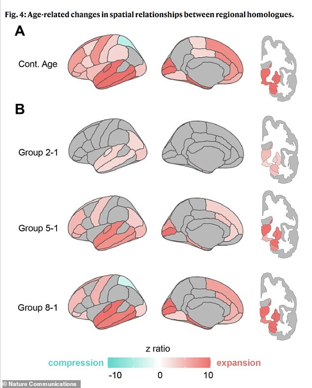
They also examined the distance between specific partner regions in the left and right hemispheres, connecting these measurements to real-world cognitive functions. Using advanced statistics, they tested whether these patterns were linked to participants’ age, memory and reasoning test performance, and whether they had been diagnosed with cognitive issues. This allowed them to see how specific changes in brain shape might affect cognitive abilities.
The aging brain undergoes a dramatic reshaping, not just shrinkage. Particular patterns of expansion and compression were strongly linked to brain health. For instance, memory problems were associated with expansion in the temporal lobe, a key area for memory. One of the study's most striking findings involves the entorhinal cortex, a vital hub for memory located in the medial temporal lobe. The research suggests that age-related brain reshaping may physically squeeze this fragile region against the base of the skull.
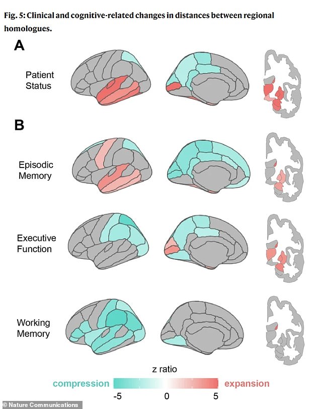
Dr. Michael Yassa, co-author of the study and a neurobiologist at the University of California, Irvine, noted that this could explain why the entorhinal cortex is a primary site for Alzheimer’s pathology. If the aging brain shifts in a way that squeezes this region against a rigid boundary, it may create conditions for damage to occur. This insight offers a new perspective on Alzheimer’s mechanisms and the possibility of early detection.
Poor executive function, including planning and reasoning, was linked to compression in parietal regions, which are involved in integrating information and vision. The patterns seen in individuals with clinical impairments were even more pronounced, suggesting that this reshaping accelerates with disease.
Janssen emphasized that this isn’t just about measuring brain shrinkage. It’s about observing how the brain’s architecture responds to aging and how that architecture predicts cognitive struggles.
The conclusion that the brain’s geometry could serve as a biomarker for dementia marks a paradigm shift in diagnosing Alzheimer’s and other forms of dementia. Their findings were published in the journal Nature Communications.
The sagging of certain brain regions could be detectable long before significant cell death occurs. A routine MRI could be analyzed to detect this, identifying a specific pattern of expansion and compression that flags a patient as high-risk years before memory tests show clear problems. Neurologists can examine a patient's brain map to identify patterns highly specific to Alzheimer’s, leading to more accurate diagnoses and targeted treatment plans.


Comments
Post a Comment